Clinical History
A ten year old, physically active boy, whose parents had enrolled him into several sports clubs, presented with limping, left perineal pain and tenderness after a soccer match. A trauma apart from normal tackling was declined by the boy, neither was it observed by his parents, who were attending.
Imaging Findings
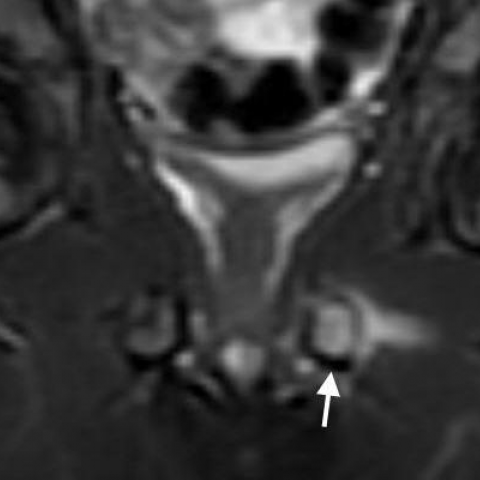
The parents went to see a pediatric orthopedist, who performed an x-ray of the pelvis and hip ultrasound. These were reportedly normal. However, images were not available to us, when the boy was referred for an MRI of the pelvis. On coronal MRI, there was T1 signal loss at the level of the left ischiopubic synchondrosis (IPS) corresponding to bright signal on T2-weighted images with fat suppression due to bone marrow oedema. The neighbouring muscles originating laterally from the inferior pubic ramus and the IPS showed circumscribed T1 and T2 hyperintense signal alterations indicative of haematoma. There were no contour defects or dislocations of the pelvic bones. On the basis of the MRI, the history, and the reported radiographic findings, the diagnosis of a stress reaction of the left IPS was made.
Discussion
The ischiopubic synchondrosis (IPS) is a temporary cartilaginous joint between the os pubis and os ischii, which is present at birth and undergoes complete ossification before puberty. A condition called ischiopubic osteochondritis was simultaneously described in 1924 by van Neck and Odelberg, based on the observation of unilateral enlargement and translucency of this joint on radiographs in symptomatic children from 5 to 10 years. These children walked with a limp and protected the affected side [1]. As to the aetiology of this condition, there has been more speculation than proof. At first, it was considered as a pathological phenomenon and designated as osteochondritis or osteochondrosis. Later, because of its apparent benignity and spontaneous healing, these terms were used less and less. Nowadays, it seems to be clear, that widening of the ischiopubic junction is a process of normal skeletal growth, at least in asymptomatic individuals. MRI is much more sensitive to certain temporary growth phenomena than conventional radiography and may therefore represent a source of diagnostic error for the inexperienced. In asymptomatic children from 4 to 16 years, who underwent pelvic MRI for other reasons, Herneth and co-workers found signal changes of the IPS in up to 86%. Hypointense fibrous bridging and fusiform swelling of the IPS were present in 68%, respectively. In more than half of cases, signal alterations of the adjacent tissues were also found [2]. These changes were observed more often unilaterally than bilaterally. This asymmetry has been attributed to increased ground reaction forces on the weight-bearing non-dominant leg, which is the left in most people [3]. Hence, mechanical stress probably does play a certain role in this condition. Although there is no proof, overuse in certain sports or recreational activities has been incessantly accused of causing a painful IPS in children of a certain age. A case of a biopsy-proven stress fracture in an eight year old boy was published only recently [4]. Although modern state-of-the-art imaging was available in this case, biopsy had to be performed out of diagnostic uncertainty. This exemplifies the diagnostic difficulty of excluding a neoplasm or haematogenic osteomyelitis in unilateral symptomatic IPS. However, in most of these rare cases, MRI can substantially increase diagnostic certainty [5]. In the present case, pain resolved spontaneously. Follow-up imaging studies were not performed. It was diagnosed as stress reaction of the IPS, formerly known as van Neck Odelberg disease.
Differential Diagnosis List
Stress reaction of the ischiopubic synchondrosis
Van Neck Odelberg disease (historical)
Osteomyelitis
Bone neoplasm
Normal skeletal development
Final Diagnosis
Stress reaction of the ischiopubic synchondrosis