Clinical History
A 10-year-old female patient presented with roncopathy, obstructive sleep apnoea and a right lateral cervical mass, which had enlarged progressively in the past four months. The patient denied pain, voice changes or dysphagia. Physical examination revealed the mass to be elastic, smooth-surfaced and mobile. No neurologic deficits were found.
Imaging Findings
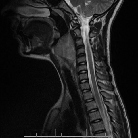
A lateral cervical spine X-ray was performed, which depicted a retropharyngeal/prevertebral mass regularly displacing the hypopharyngeal, laryngeal and upper tracheal air column anteriorly. It showed no calcifications. The cervical spine was rectified but there were no bone erosions which might suggest vertebral body involvement.
A cervical MR was performed later on. The oval-shaped solid mass was homogeneous and showed high SI on T2WI and intermediate SI on T1WI. It displaced the pre-vertebral muscles posteriorly and the hypopharynx and larynx anteriorly, suggesting a retropharyngeal origin. The oesophagus was displaced laterally to the left by the caudal portion of the mass. No clear signs of invasion of these structures were observed. There were no signs of invasion of the prevertebral muscles or cervical spine either.
No signal loss was observed on the fat-supressed images, excluding the presence of macroscopic fat. After IV contrast administration, the lesion enhanced homogeneously.
Discussion
Retropharyngeal lesions may be congenital, such as haemangioma and lymphangioma; inflammatory, either due to direct trauma (such as foreign body penetration) or due to extension of an inflammatory process from adjacent compartments (such as the pharyngeal mucosal space); and neoplastic, nodal metastases, particularly from squamous-cell carcinoma, and lymphoma being most common.
Benign fibrous soft tissue tumours are not usually included in the differential diagnosis of retropharyngeal lesions. They can be grouped in four broad categories: benign fibrous proliferations, fibromatosis, fibrosarcomas and fibrous proliferations of infancy and childhood.[1] Nodular fasciitis is included in the first group (benign fibrous proliferations) along with other well known entities, such as fibromas of the tendon sheath, keloids and hypertrophic scars, and elastofibromas.[1] It consists of a benign, tumour-like proliferation of fibroblasts [2,3] and myofibroblasts [1], and may be mistaken for a sarcomatous lesion due to its rapid growth [1,3] and pseudosarcomatous histologic features, namely high celullarity and increased mitotic activity.[3] Its pathogenesis is unknown. Some speculate that it might be reactive to trauma [1,3], while others described chromosomal abnormalities that suggest a true neoplastic origin.[1]
Nodular fasciits usually occurs between 20 and 40 years.[1,3] Fewer than 20% of lesions occur before 20 years.[3] Both sexes are affected equally.[3] It usually presents as a subcutaneous (most frequent), intramuscular or fascial, solitary [3], rapidly growing mass.[1] Nearly 50% are found in the upper extremity, anterior surface of the forearm being the most common location.[1] Head and neck lesions represent 18% of all lesions.[1] Lesions may be associated with pain and tenderness.[3]
On CT and MR, lesions are usually well-defined, less than 4cm, soft-tissue masses, but deeper, particularly intramuscular lesions, may present with a more infiltrative growth pattern, and may even invade and destroy adjacent structures, including bone.[3] MR SI is variable and results from the high variability of histologic features - some lesions are highly cellular, presenting nearly isointense to muscle on T1WI and hyperintense on T2WI, while others are more fibrous, presenting with low SI on all sequences.[1] Contrast enhancement is usually diffuse but may be peripheral when a higher extracellular myxoid component or a central cystic space is present.[1]
The differential diagnosis of nodular fasciitis includes extrabdominal desmoid tumour, fibrous histiocytoma, fibrosarcoma neurofibroma and soft-tissue sarcoma.[1,3] Intramuscular lesions may also simulate early miositis ossificans.[1]
Definite diagnosis is histologic and treatment consists of marginal excision, with a low rate of recurrence (1%).[1] Watchful waiting and steroid injection have also been advocated.[1]
Differential Diagnosis List
Retropharyngeal nodular fasciitis
Nodal metastases
Lymphoma
Final Diagnosis
Retropharyngeal nodular fasciitis