Clinical History
A 34 year old female presented to our emergency department with acute severe headache. She was agitated and confused. There was disorientation in time and space. Neurologic examination did'nt show
any focal signs. Blood pressure was normal.
Imaging Findings
A 34 year old female presented to our emergency department with acute severe headache. She was agitated and confused. There was disorientation in time and space. Neurologic examination did'nt show
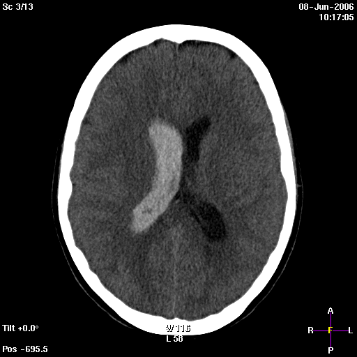
any focal signs. Blood pressure was normal. A computed tomography scan was performed which showed intraventricular hemorrhage without an evident bleeding source. A CT-angiography did not reveal an
aneurysm or a vascular malformation. A DSA followed and also did not disclose any vascular abnormality as an explanation for the intraventricular blood. Also a MRI-scan was done after 4 weeks to rule
out DSA occult vascular lesions like a cavernous angioma. The MRI-scan excluded a cavernoma but revealed volume loss of the head of the caudate nucleus compared to the left side. A meticulous look to
the initial CT-scan revealed a hypodensity at the head of the caudate nucleus compatible with an infarction. A periventricular infarction with subependymal hemorrhagic transformation was the most
probable explanation for the primary intraventricular hemorrhage.
Discussion
Primary intraventricular hemorrhage is a rare type of nontraumatic intracranial hemorraghe (2,5%) in which only blood is seen inside the ventricle. In premature infants however intraventricular
hemorrhages are common and originate in the immature periventricular germinal matrix. In infants the periventricular region is believed to represent a watershed area between the ventriculopetal and
ventriculofugal branches of the deep medullary arteries. This anatomic situation is not the same for adults where the watershed areas are believed to lie between the major vascular territories.
Secondary intraventricular hemorrhages are more prevalent and are caused by intraventricular extension from subarachnoid, traumatic or intraparenchymal bleeding. Two groups of clinical presentation
are discerned : the first group is charactarized by sudden decreased consciousness or coma and the second group is charactarized by sudden signs od increased intracranial pressure
(headache-vomiting-confusion-drowsiness). Focal signs are usually absent. An AVM can be found in circa 30% of cases as stated by Passero et al, which stresses the importance of doing a DSA. For a lot
of patients (25-48%) however a definite cause is not found. Possible risk factors mentioned in the literature are hypertension, alcoholism, tobacco, diabetes mellitus,coagulopathies and previous
strokes. Well-known causes are vascular disorders (cavernoma - AVM - aneurysms - FMD - Moya Moya) and tumors. Gates et al described an intriguing neuropathologic finding of small areas of hemorrhagic
necrosis in the wall the lateral ventricle in the caudate nucleus as a possible explanation for primary intraventricular hemorrhages. We present a similar case in which primary intraventicular
hemorrhage occured in conjunction with a hypodense, ischemic lesion in the head of the caudate nucleus. Our case demonstrates intraventricular hemorrhage can occur without parenchymal hemorraghic
transformation on imaging studies. The amount of intraventricular blood as well as the presence of early hydrocephalus are important prognostic factors. Hydrocephalus is the most important
complication followed by re-bleeding. Mortality rate approximates 50%, those who survive usually don't have any physical disability, nevertheless they frequently suffer from cognitive dysfunction.
Differential Diagnosis List
primary intraventricular hemorrhage caused by ischemia in the caudate nucleus
Final Diagnosis
primary intraventricular hemorrhage caused by ischemia in the caudate nucleus