Figure 1
Fig 1a: a draw of the hemisphere’s lateral face. 1: frontal lobe; 2: parietal lobe; 3: occipital lobe; 4: temporal lobe; 5: insula lobe. (4)

Neuroradiology
Case TypeAnatomy and Functional Imaging
AuthorsDr. I. Trivelli
33 years, female
[1]
1. C. Balboni.
Anatomia Umana
Ed. Ermes (1993).
[2]
2. G. Simonetti, A. Del Maschio, C. Bartolozzi, R. Passariello.
Trattato italiano di Risonanza Magnetica.
Ed. Idelson-Gnocchi (1998).
[3]
3. Duane E. Haines.
Neuroanatomy
Lippincott Williams and Wilkins (2000).
[4]
4. Interbrain
Springer-Verlag
Electronic Media Dept. April 1998.
| URL: | https://www.eurorad.org/case/3541 |
| DOI: | 10.1594/EURORAD/CASE.3541 |
| ISSN: | 1563-4086 |
Fig 1a: a draw of the hemisphere’s lateral face. 1: frontal lobe; 2: parietal lobe; 3: occipital lobe; 4: temporal lobe; 5: insula lobe. (4)
Fig 1b: a draw of the hemisphere’s medial face. 1: frontal lobe; 2: parietal lobe; 3: occipital lobe; 4: temporal lobe; 5: limbic lobe. (4)
Fig 2a: T1 MRI axial projection. 1: inter-hemispheric scissure; 2: lateral sulcus; 3: frontal lobe; 4: insula lobe; 5: temporal lobe; 6: occipital lobe.
Fig 2b. 1: frontal pole; 2: frontal lobe; 3: parietal lobe; 4: inter-hemispheric scissure; 5: central sulcus; 6: cingulate sulcus.
Fig 2c. 1: falx cerebri; 2: frontal lobe; 3: central sulcus; 4: parietal lobe.
Fig 3a: Inverted T1 MRI axial projection. 1: frontal lobe; 2: temporal lobe; 3: lateral sulcus; 4: cingolate gyrus; 5: parietal lobe.
Fig 3b. 1: inter-hemispheric scissure; 2: frontal lobe; 3: central sulcus; 4: parietal lobe; 5: cingolate gyrus.
Fig 4a: Inverted T1 MRI coronal projection. 1: inter-hemispheric scissure; 2: lateral sulcus; 3: insula lobe; 4: frontal superior gyrus; 5: frontal middle gyrus; 6: frontal inferior gyrus; 7: temporal pole
Fig 4b. 1: inter-hemispheric scissure; 2: lateral sulcus; 3: deep recess of the lateral sulcus; 4: frontal superior gyrus; 5: frontal middle gyrus; 6: frontal inferior gyrus; 7: temporal pole.
Fig 5a: T1 MRI coronal projection. 1: inter-hemispheric scissure; 2: lateral sulcus; 3: temporal lobe; 4: frontal superior gyrus; 5: frontal middle gyrus; 6: frontal inferior gyrus.
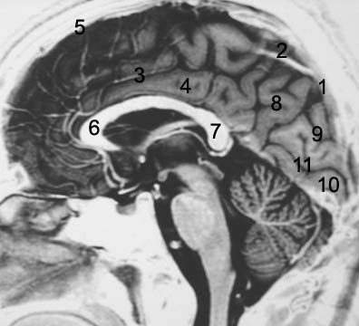
Fig 6a: Inverted T1 MRI sagittal projection. 1: parietoccipital sulcus; 2: central sulcus; 3: limbic (cingolate) sulcus; 4: limbic lobe; 5: frontal lobe; 6: corpus callosum rostro; 7: corpus callosum spenio; 8: parietal lobe; 9: occipital lobe (cuneo region); 10: occipital lobe (lingual lobe); 11: calcarine sulcus.
Fig 6b. 1: parietoccipital sulcus; 2: central sulcus; 3: frontal pole; 4: frontal lobe; 5: parietal lobe; 6: occipital lobe (cuneo region); 7: calcarine sulcus; 8: occipital lobe (lingual lobe).
Fig 6c. 1: parietoccipital sulcus; 2: central sulcus; 3: frontal lobe; 4: parietal lobe; 5: occipital lobe; 6: cerebellum.