Background
(definitions, disease description, pathophysiology)
Renal-tract fistula is a recognised phenomenon originating either from trauma or spontaneously through chronic inflammatory states in the kidney, commonly as a result of calculi, infection and malignancy.
Reno-duodenal fistulae are rare comprising 1% of renoalimentary fistulae [1]. The main side involved is the right one; this is due to the proximity of right kidney to the descending duodenum with its relative immobility, lack of posterior peritoneal covering, and close contact with the anterior kidney [2]. Lin et al. demonstrated that 58.9% of the renoalimentary fistula are renocolic, 34.8% renoduodenal and the remaing 6.3% renojejunal or renoileal. Pionephrosis appears to be the most common cause of renoduodenal fistula (25.5%), followed by complicated nephrolithiasis (18.2%), iatrogenic causes (10.9%), malignancy and GI causes (9.1%), infectious disease and trauma (7.3%), and xanthogranulomatous pyelonephritis (5.5%) [3].
Imaging Perspective
(diagnostic pearls, key findings, which diagnostic procedures are useful, how is the final diagnosis made)
Typically, CT scanning identify renal-fistula, but there are other imaging modalities, like retrograde pyelography, oesophago-gastro-duodenoscopy (OGD) and 99mTc scintigraphy that can be helpful in the diagnosis. [4]
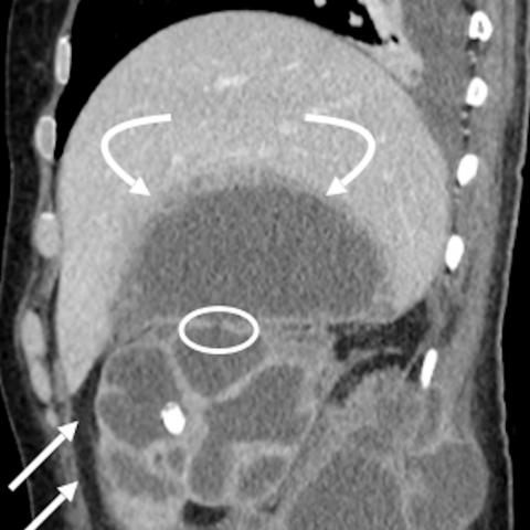
On CT, inflammatory changes and/or adhesion between the kidney and the gastrointestinal tract may suggest the diagnosis of reno-alimentary fistula, particularly if we find, like in this case, calculi in the via fistulous. Obviously, it is necessary to find other causes that are responsible of the pathology, like chronic perinephric inflammation, renal calculi and/or obstruction, tumours, surgical instrumentation of the genitourinary tract, penetrating trauma, diseases in the gastrointestinal tract and others.
Outcome
(therapeutic options, prognosis, impact of imaging on therapy planning)
Depending on the renal function, there are three main treatments: open nephrectomy with duodenal oversewn or IV antibiotics with urinary diversion through either ureteric stent insertion or nephrostomy [4] or endoscopic treatment by application of clips via OGD to close the fistula tract and subsequent ligation of the fistula tract using an endoloop [5].
Take-Home Message / Teaching Points
This case demonstrates a rare complication of renal tract calculi along with other chronic inflammatory processes within the kidney and highlights the role of CT for the right diagnosis and early treatment.
Written informed patient consent for publication has been obtained.