


Figure 1
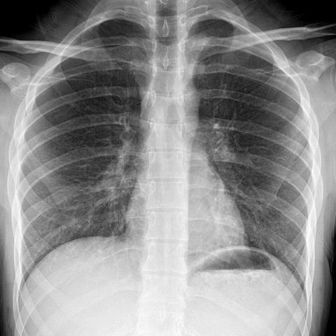
Chest X-Ray; Apparently Normal Chest X-Ray Study
Chest imaging
Case TypeClinical Cases
Authors
Akin Cinkooglu MD*, Habib Ahmad Esmat MD*, Savas Recep MD, Mohammad Naim Forogh MD,
19 years, male
A 19-year-old male presented to our hospital with fever and cough. No history of traveling or other comorbidity. The Chest X-Ray was unremarkable (Fig.1), but, on his chest CT, a small single ground-glass opacity in the left upper lobe of the lung was observed (Figs. 2a, 2b, 2c). His real-time PCR was positive for COVID-19.
In the left upper lobe of the lung, a small area of ground-glass opacity was observed, however, both lungs aeration and vascularization were within normal limits. Pleura and interlobar fissures were normal.
Background
The initial cases of pneumonia of unknown aetiology first identified on Dec 31, 2019, by the Health Commission of Hubei province of China. [1]. On February 11, 2020, the World Health Organization named this disease as Corona Virus 2019 (COVID-19) [2]. The virus is also called as SARS-Cov2, which is a class of enveloped, positive-sense, single-strand RNA viruses that can cause respiratory, enteric, hepatic, and neurologic diseases [3]. It is mainly transmitted through nosocomial transmission, respiratory droplets, and contact [4]. Studies support the COVID-19 S-protein interaction with human ACE2 receptors in the pathogenesis of COVID -19, leading to cellular entry [5].
Clinical Perspective
The common symptoms of disease are fever, cough, myalgia, fatigue, and dyspnoea [6]. Although the real-time reverse-transcription-polymerase chain reaction (RT-PCR) is the standard of diagnosis, imaging can play a useful complementary role in determination and management of COVID-19 pneumonia [7].
Imaging Perspective
Bilateral multifocal ground-glass opacities (GGO) in the lower lobes are the most common initial findings on CT [8]. Pleural thickening, bronchiectasis, and septal thickening are less common imaging manifestations and mainly seen in the later stages [9]. The PCR is the reference standard for the diagnosis of COVID-19 but, in some cases of the early stages of diseases, it may have a false-negative result [10]. Thus, in patients with typical clinical features, chest CT may play a useful complementary role in the integrated diagnosis of disease even when the PCR result is negative [11].
Outcome
COVID-19 pneumonia has nonspecific and diverse CT imaging findings but, GGO is the most typical feature. [12]. In patients with typical clinical features and high suspension, chest CT may play a useful complementary role in disease diagnosis and management, especially where the PCR access is difficult [7, 11].
Take-Home Message
Although the COVID-19 pneumonia often manifests as multifocal GGO with peripheral distribution in the lower lobes it can be presented as a single GGO in the upper lobe.
[1] Chan JF, Yuan S, Kok KH, et al (2020) .A familial cluster of pneumonia associated with the 2019 novel coronavirus indicating person-to-person transmission: a study of a family cluster. The Lancet: 395 (10223), 514-523 .20 30154-9
[2] Hussin A. RothanSiddappa N. Byrareddy (2020) .The epidemiology and pathogenesis of coronavirus disease (COVID-19) outbreak. Journal of Autoimmunity : 109: 102433.102433
[3] Jin, Y,et al (2020). Epidemiology, Pathogenesis, and Control of COVID-19. Viruses , 12, (4),372 .12040372
[4] Han Y,Yang H(2020). The transmission and diagnosis of 2019 novel coronavirus infection disease (COVID -19): A Chines Perspective. J MED Virology :92: 639-644. 25749
[5] Guo YR, Cao QD, Hong ZS, et al(2020). The origin, transmission and clinical therapies on coronavirus disease 2019 (COVID-19) outbreak - an update on the status. Mil Med Res:7(1):11. 40779-020-00240-0
[6] Adhikari, S., Meng, S., Wu, Y. et al. (2020).Epidemiology, causes, clinical manifestation and diagnosis, prevention and control of coronavirus disease (COVID-19) during the early outbreak period: a scoping review. Infect Dis Poverty 9, 29 . 40249-020-00646-x
[7] Sverzellati N, Milone F, Balbi M(2020). How imaging should properly be used in COVID-19 outbreak: an Italian experience: Diagn Interv Radiol . 2020.30320.
[8] Xu,X et al (2020).Imaging and clinical features of patients with 2019 novel coronavirus SARS-CoV-2: Eur J Nucl Med Imaging.47,1275-1280.0059-020-04735-9
[9] Sana Salehi, et al(2020).Coronavirus Disease 2019 (COVID-19): A Systematic Review of Imaging Findings in 919 Patients: American Journal of Roentgenology,14:1-7.20.23034.
[10] Xie X, Zhong Z, Zhao W, Zheng C, Wang F, Liu J. Chest CT for typical 2019-nCoV pneumonia :relationship to negative RT-PCR testing. Radiology (2020) .2020200343
[11] Kaiyue Diao,Peilun Han, et al .(2020) HRCT imaging features in representative imported cases of 2019 novel coronavirus pneumonia:Precision Clinical Medicine.3, 9-13.doi.org/10.1039/pcmeddi/pbaa004
[12] Bingjie Li, Xin Li, Yaxuan Wang et al.(2020).Diagnostic value an key features of computed tomography in Coronavirus Disease 2019, Emerging Microbes & Infection,9:1,787-793. DOI.org/10.1080/22221751.2020.1750307
| URL: | https://www.eurorad.org/case/16903 |
| DOI: | 10.35100/eurorad/case.16903 |
| ISSN: | 1563-4086 |
This work is licensed under a Creative Commons Attribution-NonCommercial-ShareAlike 4.0 International License.


