


Figure 1
Chest X-Ray, AP and lateral
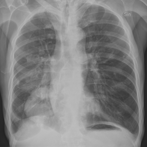
A big rounded tumor in the right lower lobe with cavitation. Calcified pleural plaque bilaterally and findings of COPD.
Chest imaging
Case TypeClinical Cases
Authors
Gracijela Bozovic
79 years, male
An elderly gentleman of 79 years suffering from diabetes and COPD with asbestos exposure and smoking history, and previous malignant melanoma from which he had been declared healthy. He complains about weight loss and declining wellbeing. He had palpable cervical lymph nodes and pain in one rib dorsolateral on the right side.
Figure 1 and 2. Chest X-Ray, AP and lateral
A big rounded tumour in the right lower lobe with cavitation. Calcified pleural plaque bilaterally and findings of COPD.
Figure 3 and 4. CT Chest with iv contrast
Subpleural round tumour measuring 8 cm in diameter in the right lower lobe with well-defined borders and parts of low attenuation and cavitation in keeping with necrosis. Pleural involvement with a small amount of right-sided pleural effusion. Enlarged lymph nodes in the right hilum and mediastinum and metastasis with bone destruction in costa X on the right side. Bilateral calcified plaque with distribution suggesting asbestos exposure. Moderate emphysema.
Figure 5 and 6.
Papanicolaou staining of cytological material from endobronchial ultrasound-guided fine-needle aspiration showing keratinising squamous cell carcinoma.
Figure 7, 8 and 9. CT Chest with iv contrast performed three years previously
A round atelectasis in the right lower lobe where the tumour is present now. It is defined by its subpleural localisation adjacent to thickened pleura, wedge-shaped mass and curvilinear vessels and bronchi entering the mass (“comet sign”).
Round atelectasis (RA) also known as folded lung, Blesovsky syndrome or atelectatic pseudotumour is a form of chronic pleuro-pulmonary disease. It emerges due to pleural disease with thickening, usually pleural fibrosis causing adhesions to the adjacent lung. The lung is trapped and over time gradually folded, probably facilitated by retraction of the pleura [1]. There are certain imaging findings such as subpleural location, pleural thickening of the adjacent pleura (88%), converging bronchovascular markings extending towards the hilum, associated ipsilateral pleural fluid (60%), and small calcifications (32%), more commonly men (88%) and most often found in the lower lobes [1,2]. In a series of 50 patients, the follow up stretching to 126 months 88% stayed stable or decreased in size (88%) [2]. If up filling the criteria the diagnosis of RA is straight forward. Sometimes there is a need for verification with a biopsy. On PET it shows increased uptake making it difficult to differ from malignancy.
RA is commonly associated with asbestos-related pleural disease but has more often other causes such as infections, uremic or congestive heart disease pleuritic effusion and benign pleural exudates e.g. as in congestive heart disease (25% are exudate) or systemic diseases with serositis [3]. They can be associated with mesothelioma if asbestos-related. Lung cancer can also occur in RA although very rare since there are only two reported cases, one of them probably being coincidental [4]. It is unknown if RA itself is a risk factor for developing lung cancer or if it is coincidental. The coexistence is rare [5] and if present other risk factors such as smoking and asbestos exposure are more significant [6]. Given the rareness, RA as a risk factor itself is not likely. Nonetheless, the finding of a RA should always be put in a clinical context and preferably followed up. If any diagnostic doubt extensive investigation is mandatory. Regarding the timeline for the follow up, there are no recommendations.
This patient developed a squamous cell lung carcinoma in a RA. Unfortunately, he fell out from the follow up after the first chest CT and returned with advanced lung cancer and limited treatment options. He passed away within months of the diagnosis. This would be the third reported case with lung cancer in a RA.
[1] David Hansell, David Lynch H, Page McAdams, Alexander Bankier. Imaging of Diseases of the Chest 5th Edition. Mosby. December 2009.
[2] Azour L, Billah T, Salvatore MM et al. Causative factors, imaging findings, and CT course of round atelectasis. Clinical Imaging 50 (2018) 250–257. PMID: 29704809
[3] Stathopoulos GT, Karamessini M, Sotiriadi AE et al. Rounded atelectasis of the lung. Respiratory Medicine (2005) 99, 615–623. PMID: 15823460
[4] Temes E, Noya A and Troncoso A. Lung Cancer Coexisting in Round Atelectasis. Arch Bronconeumol 2004;40(7):333-5.
[5] Fraser RS, Muller NL, Colman N, Pare PD. Diagnosis of diseases of the chest, 4th ed. Philadelphia, PA: W. B. Saunders, 1999:521–522
[6] Malhotra J, Malyazzi M, Neggri E et al. Risk factors for luncancer worldwide. European Respiratory Journal 2016:48:889-902.
| URL: | https://www.eurorad.org/case/16798 |
| DOI: | 10.35100/eurorad/case.16798 |
| ISSN: | 1563-4086 |
This work is licensed under a Creative Commons Attribution-NonCommercial-ShareAlike 4.0 International License.

Chest X-Ray, AP and lateral
Chest X-Ray, AP and lateral
CT Chest with iv contrast
CT Chest with iv contrast
Ultrasound-guided fine-needle aspiration
Ultrasound-guided fine-needle aspiration
CT Chest with iv contrast performed three years previously
CT Chest with iv contrast performed three years previously
CT Chest with iv contrast performed three years previously


