Periarticular ossification and myositis ossificans could appear after a major trauma such as surgery for hip prosthesis or extended burn [2] and present typical bone organisation with a distinct cortical and trabecular bone pattern along with calcifications. Calcifications normally appear as mineralised densities with an attenuation coefficient higher than soft tissue but lower than bone [1].
Calcifications may arise in septic arthritis, crystal deposition diseases or acute calcific periarthritis (ACP) [3]. For septic arthritis, calcifications are a late manifestation and clinical manifestations are different (blush, heat, fever, etc.) [3]. In pseudogout, calcifications may present with acute symptoms and similar-appearing calcifications, but it is unusual to identify those calcifications without associated chondrocalcinosis. In gout, calcifications are only seen in the intermediate and late stages of chronic gout and are associated with erosions and cortical irregularities. Calcifications are typically associated with a soft-tissue tophus. During intermediate stages, joint space loss may also be seen. Patients have a history of recurrent attacks [3].
In basic phosphocalcic crystal deposition disease, mostly hydroxyapatite, calcifications may appear in either the periarticular tissues or in the intra-articular space. These calcifications are amorphous, dense, round or oval and may have a cloudy aspect while resorbing [4]. For typical phosphocalcic deposits in the supraspinous tendon the period of resorption of the calcifications is usually very painful, which is not the case for our patient. ACP must be considered a subset of hydroxyapatite deposition disease [3]. Calcifications of ACP appear without a triggering factor but early on in the disease and then are gradually adsorbed only with symptomatic treatment and splinting. Radiographs show dense, homogeneous, amorphous, cloud-like, generally round or oval calcific deposits with no cortex or internal trabecular pattern [3], periarticular, with no evidence of joint space narrowing or cortical lesion. Deposit may remain static for a long period, enlarge and change shape, or diminish in size and disappear. Recurrence is not usual for ACP [5].
We also should consider calcifications in systemic processes but where clinical presentation is different [3].
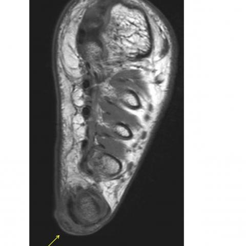
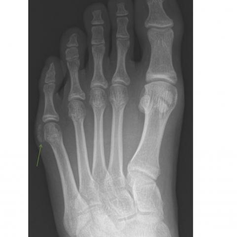
In our case, MRI excluded a tumoral lesion. Recurrent painful swelling when mechanical conflict exists without other symptoms rules out septic arthritis, crystal deposition diseases and systemic processes. The seasonal evolution of symptoms, association with a quintus varus and the absence of recurrence after surgery are in favour of a micro-traumatic origin associated with mechanical conflict.
Written informed patient consent for publication has been obtained.