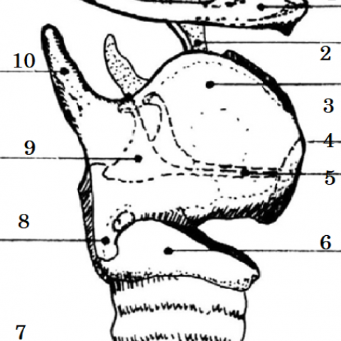
Side view of laryngeal skeleton

Head & neck imaging
Case TypeClinical Cases
Authors
Micheline Soro Tiona, Omar Kacimi, Najwa Touil, Houria Tabakh, Nabil Chikhaoui
Patient39 years, male
A 39-year-old man who presented with thoracic pain and dysphonia after a highway accident. Case history and physical examination revealed thoracic impact with the steering wheel. He was addressed for cervico-thoracic CT.
Computed tomography (without contrast material injection) of the cervico-thoracic region revealed cricoid cartilage fractures complicated by rupture of the first tracheal ring. The lesion presented as a discontinuity of cricoid cartilage with displacement of fracture fragments. These fractures were associated with soft-tissue haematoma, and extensive emphysema extending to anterosuperior mediastinum. The analysis of the trachea notes a deformation of the first tracheal ring, leading us to make the diagnosis of partial fracture of trachea.
Larynx is a duct consisting of a cartilaginous skeleton that comprises three unpaired cartilages (thyroid, cricoid and epiglottis) and three smaller paired cartilages (arytenoid, cuneiform and corniculate cartilages), all connected by membranes, ligaments and muscles.
Laryngeal fracture is a rare phenomen occuring in less than 1% cases of blunt trauma [1,2].
Laryngeal fractures interest in order of frequency the thyroid, cricoid and arythenoid cartilages and are more common in older persons because of calcification of the laryngeal cartilages [3,4].
Three clinical signs indicating laryngeal fractures were proposed by the American College of Surgeon's Advanced Trauma Life Support protocol: hoarseness, subcutaneous emphysema and palpable fracture [5].
Cervico-thoracic CT plays a key role in the management of the patient by demonstrating the fracture and associated injuries like vertebral lesions.
2D and 3D multiplanar reconstructions makes it easier to detect occult fracture and misalignment in ossified cartilage [6].
Fractures of the cricoid cartilage are often bilateral [6,7,11].
Unlike the thyroid cartilage, the cricoid is poorly ossified, and these fractures may be overlooked at MDCT. It is therefore essential to analyse the soft part window or to perform an MRI in high suspicion.
Fractures appear as discontinuities of the cartilage, with or without displacement.
Partial or complete laryngo-tracheal separation may occur in up to 50% of cricoid fractures [8,9] This rupture is typically seen at the level of the first tracheal ring.
Complete rupture : the trachea retracts into the mediastinum [9]. There is widening of the crico-tracheal distance due to retraction of the trachea, and the endotracheal tube lies in an amorphous cavity caused by rupture of the crico-tracheal membrane. Subcutaneous emphysema without major pneumothorax is also commonly observed [9].
Partial transection : tracheal deformation and discontinuity, mucosal laceration, and emphysema are observed [10,11].
Endoscopy allows exploration of the mucosa and vocal cords.
At the end, the clinical, endoscopic and radiological evaluation makes it possible to classify the traumatism according to severity (classification of Schaefer and Fuhrman) [1].
Treatment of the fracture is surgical with post-operative intubation to avoid the major complication, which is tracheal stenosis [12].
Imaging plays a major role in identifying laryngo-tracheal injuries.
CT is the modality of choice for fracture detection, the soft-tissue windowing being best suited for non-ossified cartilage. An MRI will be performed in case of strong clinical suspicion when the CT is normal.
The gas bubble sign-a reliable indicator of fracture[3].
Written informed patient consent for publication has been obtained.
[1] Fuhrman GM, Stieg FH 3rd, Buerk CA. Blunt laryngeal trauma: classification and management protocol. J Trauma. 1990 Jan. 30(1):87-92. [Medline] (PMID: 2296072)
[2] Chitose, S., Sato, K., Nakazono, H., Fukahori, M., Umeno, H., & Nakashima, T. (2014). Surgical management for isolated cricoid fracture causing arytenoid immobility. Auris Nasus Larynx, 41(2), 225–228. doi:10.1016/j.anl.2013.10.013. (PMID: 24268328).
[3] Schulze K, Ebert LC, Ruder TD, et al. The gas bubble sign-a reliable indicator of laryngeal fractures in hanging on post-mortem CT. Br J Radiol. 2018 Apr. 91 (1084):20170479. [Medline] (importance de la mise en évidence d’air). (PMID: 29327945)
[4] Schaefer SD, Close LG. Acute management of laryngeal trauma. Update. Ann Otol Rhinol Laryngol. 1989 Feb. 98(2):98-104. (PMID: 2916832)
[5] Oh, J. H., Min, H. S., Park, T. U., Lee, S. J., & Kim, S. E. (2007). Isolated cricoid fracture associated with blunt neck trauma. Emergency Medicine Journal, 24(7), 505–506. doi:10.1136/emj.2007.048355. (PMID: 17582051)
[6] Becker, M., Duboé, P.-O., Platon, A., Kohler, R., Tasu, J.-P., Becker, C. D., & Poletti, P.-A. (2013). MDCT in the Assessment of Laryngeal Trauma: Value of 2D Multiplanar and 3D Reconstructions. American Journal of Roentgenology, 201(4), W639–W647. doi:10.2214/ajr.12.9813 (PMID: 24059404)
[7] Lee WT, Eliashar R, Eliachar I. Acute external laryngotracheal trauma: diagnosis and management. Ear Nose Throat J. 2006 Mar;85(3):179-84. (PMID: 16615601)
[8] Couraud L, Velly JF, Martigne C, N’Diaye M. Post traumatic disruption of the laryngo-tracheal junction. Eur J Cardiothorac Surg 1989;3(5):441–4.
[9] Becker, M., Leuchter, I., Platon, A., Becker, C. D., Dulguerov, P., & Varoquaux, A. (2014). Imaging of laryngeal trauma. European Journal of Radiology, 83(1), 142–154. doi:10.1016/j.ejrad.2013.10.021. (PMID: 24238937)
[10] Scaglione M, Romano S, Pinto A, Sparano A, Scialpi M, Rotondo A. Acute tracheobronchial injuries: impact of imaging on diagnosis and management implications. Eur J Radiol 2006;59(3):336–43. (PMID: 16782296)
[11] Steenburg SD, Sliker CW, Shanmuganathan K, Siegel EL. Imaging evaluation of penetrating neck injuries. Radiographics 2010;30(4):869–86. (PMID: 20631357)
[12] 1Bell RB, Osborn T, Dierks EJ, Potter BE, Long WB. Management of penetrating neck injuries: a new paradigm for civilian trauma. J Oral Maxillofac Surg 2007;65(4):691–705.
| URL: | https://www.eurorad.org/case/16534 |
| DOI: | 10.35100/eurorad/case.16534 |
| ISSN: | 1563-4086 |
This work is licensed under a Creative Commons Attribution-NonCommercial-ShareAlike 4.0 International License.