CASE 15636 Published on 23.04.2018

Onset of sarcoidosis with predominant liver manifestations in a young Caucasian man

Section

Abdominal imaging

Case Type

Clinical Cases

Authors

Galateia Skouroumouni, Charikleia Mauridou, Despina Panagiotidou, Melpomeni Kosmidou, Stavroula Pervana, Sofia Papaioannou

Papageorgiou General Hospital,
Radiology;
Pavlos Melas Street
56429 Thessaloniki, Greece;
Email:galskour@hotmail.com
Patient

35 years, male

Categories
Area of Interest Liver, Lymph nodes, Lung ; Imaging Technique CT, MR, Experimental
Clinical History
A 35-year-old Caucasian male patient, without significant medical history, presented with longstanding abdominal pain. Abdominal examination showed hepatomegaly. Laboratory tests revealed elevated liver enzyme levels (alanine transaminase, 82 IU/l; aspartate transaminase, 68 IU/l; γ-glutamyltranspeptidase, 258 IU/l; alkaline phosphatase, 646 IU/l). Testing of possible infectious, autoimmune and malignant liver processes was negative.
Imaging Findings
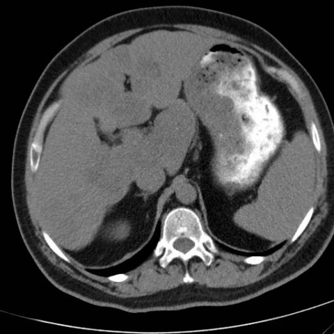
Non-enhanced abdominal CT (Fig. 1) showed cirrhotic changes of the liver, with increased size of the left and caudate lobe, contour irregularity, and parenchymal heterogeneity. Contrast-enhanced abdominal CT (Fig. 1) revealed a pattern of multiple hypodense macro-nodules. Signs of portal hypertension or involvement of the spleen were absent. Abdominal MRI (Fig. 2) was scheduled. T1-weighted MR-images showed large confluent hypointense nodules. Nodules appeared slightly hyperintense in T2-weighted MR-images, hypointense on T2-weighted fat-saturated MR-images, and demonstrated signal dropout on opposed-phase images. Abdominal lymph nodes were not enlarged. No foci of abnormal enhancement or restricted diffusion were detected. Vascular architecture and bile duct morphology were intact. A liver biopsy was performed, revealing non-caseating epithelioid granulomas, large regenerative nodules with fat deposition and fibrotic changes (Fig. 3). A subsequent chest CT (Fig. 4) revealed mild hilar and mediastinal lymphadenopathy. Intrapulmonary nodules were also seen. Resolution of chest imaging findings was noted after 6 months of treatment with corticosteroids and azatheioprine.
Discussion
Sarcoidosis is a multi-systemic non-caseous granulomas disease of unknown origin that commonly affects the liver; reported series show 50-80% of patients with liver involvement, based on biopsy results.[1] Hepatic sarcoidosis is mostly clinically silent. Only a minority of patients can have a severe and rapid progression, with the occurrence of complications, including cirrhosis, portal hypertension, and chronic cholestatic disease.[2] The pathophysiology of cirrhosis due to sarcoidosis is not clearly reported. However, it has been suggested that confluent granulomas along the periportal track may ultimately lead to fibrosis with cirrhotic remodelling of the liver.[3]

The most common abnormal presentation of liver function tests in hepatic sarcoidosis is an elevation in serum alkanine phosphatase level, which can be elevated 5-10 times the upper limit of normal. The severity of liver test abnormalities is directly associated with the extend of the granulomatous inflammation.[4]

The imaging findings of hepatosplenic sarcoidosis are usually not specific (hepatomegaly, splenomegaly, cirrhotic changes). Histologically, granulomatous hepatic lesions are generally less than 2 mm in size.[5] Imaging studies depict nodular changes in only a minority of patients. On imaging, size of the lesions is reported between 2 mm and 15 mm.[5, 6] Sarcoid nodules appear hypodense at CT, with decreased vascularity.[7] At ultrasound examination, larger granulomas can be seen as hypoechoic nodules. At MRI, the lesions are hypointense on T2-weighted sequences, especially in T2-weighted fat-saturated sequences, and on gadolinium-enhanced T1-weighted sequences. However, nodules with active inflammation can appear hyperintense on T2-weighted images and demonstrate restricted diffusion.[8] In our case, large nodules represented regenerative nodules on the basis of liver cirrhosis, whereas sarcoid nodules were not apparent on imaging studies (CT, MRI).

Macro-nodular cirrhosis in a patient, with no history of alcohol abuse or viral hepatitis, have to be differentiated from other entities, such as non-alcoholic steatohepatitis(NASH). Synchronous appearance of sarcoidosis and NASH has, to our knowledge, never been reported. Liver cirrhosis due to sarcoidosis is rare, but should be considered in the differential diagnosis of cryptogenic cirrhosis. Moreover, metastases and lymphoma had to be excluded, in respect to the differential diagnosis of large atypical hepatic nodules.

The diagnosis of hepatic sarcoidosis is clinically and radiologically challenging. The diagnosis is made by clinical and radiologic findings suggestive of sarcoidosis, supported by histopathologic findings of non-caseating granulomas on biopsy, after the exclusion of other causes of hepatic granuloma formation.

Evidence based guidelines for the treatment of hepatic sarcoidosis are lacking.
Differential Diagnosis List
Macro-nodular liver cirrhosis due to sarcoidosis
Non-alcoholic steatohepatitis-associated cirrhosis
Alcoholic liver cirrhosis
Viral hepatitis-associated cirrhosis
Lymphoma
Metastasis
Final Diagnosis
Macro-nodular liver cirrhosis due to sarcoidosis
Case information
URL: https://www.eurorad.org/case/15636
DOI: 10.1594/EURORAD/CASE.15636
ISSN: 1563-4086
License