Clinical History
An 86-year-old patient presented with fever, disorientation and urinary incontinence. Urinary cultures were positive, and the patient was diagnosed with urinary tract infection. His condition worsened three days after antibiotic treatment, and abdominal CT was performed to exclude complications.
Imaging Findings
Contrast-enhanced CT (CECT) showed a fluid collection lateral to the bladder and with fat stranding (Figure 1a, b, c). An enhancing tubular connection between the bladder and the collection was present (Figure 1d).
On axial (Figure 2a) and sagittal (Figure 2b) CECT performed one week later, a rounded lateral bladder diverticulum was seen. Coronal CT image displayed the diverticulum at the left lateral aspect of the bladder (Figure 2c). The stalk of the diverticulum was visible between the bladder and the diverticulum (Figure 2d).
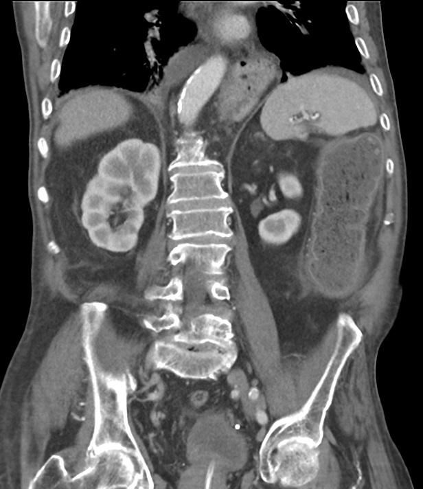
In addition, the axial (Figure 3a) and coronal (Figure 3b) CECT showed a stenotic sigmoid wall narrowing with dilation and wall thickening of proximal bowel loops. Distention and wall thickening of transverse and descending colon was identified on coronal (Figure 3c) and sagittal (Figure 3d) CECT.
Discussion
Bladder diverticula are evaginations from the bladder mucosa. In general, bladder diverticula are seen in imaging studies for urinary tract infection, incontinence or retention.
Bladder diverticula may be classified as congenital and acquired. Acquired diverticula are commonly multiple, smaller in size and usually result from chronic lower urinary tract obstruction. Congenital diverticula have been described to be associated with Ehlers-Danlos or Prune-Belly syndromes.
Bladder diverticula may be associated with a range of complications including rupture, stones and cancer. There is a well-established relationship between urinary bladder diverticula and intradiverticular urothelial carcinoma.
In younger patients, ultrasound is preferred to evaluate the presence of bladder diverticula. Occasionally, Doppler US proves to be useful to distinguish between a clot and neoplasia.
CT is considered a reliable method for the diagnosis of bladder diverticula. Furthermore, CT allows the detection of associated lesions in- and outside the urinary tract.
In this patient, CT enabled the diagnosis of cystitis, and a giant collapsed bladder diverticulum with a typical diverticular neck confirming its origin from the bladder.
As the patient's condition worsened, a repeat CT examination was performed one week later now showing a neoplasm of the sigmoid colon. In retrospect, the bowel neoplasm was visible at the first CT examination.
The case illustrates the satisfaction of search error, meaning the failure to detect a subsequent abnormality or abnormalities after an initial one has been observed.
Differential Diagnosis List
Cystitis with bladder diverticulum. Stenotic sigmoid neoplasm and bowel obstruction.
Paravesical abscess
Bowel Ischaemia
Final Diagnosis
Cystitis with bladder diverticulum. Stenotic sigmoid neoplasm and bowel obstruction.