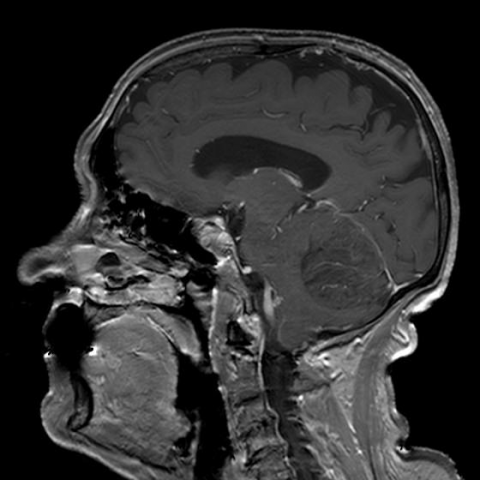
CASE 14955 Published on 24.08.2017

Dysplastic gangliocytoma of the cerebellum in an elderly patient: MR-imaging

Section

Neuroradiology

Case Type

Clinical Cases

Authors

Katerina Manavi, Galateia Skouroumouni, Filippos Sarafis, Giannis Petmezaris, Eliza Stavride, Ioannis Tsitouridis

Papageorgiou General Hospital,
Radiology;
Voulgaroktonou 4A 5
5535 Thessaloniki, Greece;
Email:galskour@hotmail.com
Patient

77 years, female

Categories
Area of Interest Neuroradiology brain ; Imaging Technique MR, MR-Diffusion/Perfusion
Clinical History
A 77-year old woman presented to our institution with persistent headache, gait instability, dysarthria and dysphagia. A head MRI was scheduled.
Imaging Findings
MR imaging of the head revealed a large lesion occupying nearly the entire left cerebellar hemisphere, extending into the vermis, compressing the fourth ventricle and causing mild obstructive hydrocephalus. There was no cerebellar herniation and the temporal horns were not remarkably dilated. The lesion was hypointense on T1W images and hyperintense on T2W images, with parallel linear striations which resulted in a "tiger striped" appearance of the involved cerebellar surface (Figure 1). Gadolinium-enhanced images showed no enhancement (Fig. 2). The abnormal area was seen with high signal on isotropic diffusion-weighted images, while in the apparent diffusion coefficient (ADC) map the mass appeared isointense to the cerebellar parenchyma (Fig. 3). The pathognomonic MRI findings undoubtedly indicated the diagnosis of dysplastic gangliocytoma and considering the patient’s old age no intervening procedures were performed.
Discussion
Lhermitte-Duclos disease (LDD) is defined as dysplastic cerebellar gangliocytoma and categorized as a WHO grade I tumour. [1] LDD is a rare, slow-growing, progressive, non-malignant tumour which presents as a mass in the posterior cranial fossa. It often becomes clinically apparent in the third to fourth decade of life. [2, 3]

LDD is characterised by progressive enlargement of the cerebellar cortex. [4] The histological findings typically show non-neoplastic tissue consisting of cerebellar folia occupied by hypertrophic neurons of the internal granule cell layer. [5] The outer layer is dominated by hypermyelinated axons from abnormal ganglion cells. Mitosis and proliferation are absent, and Purkinje cells and white matter are diminished.

Clinical symptoms are caused by gradual increase of intracranial pressure (mass effect/secondary obstructive hydrocephalus), and cerebellar dysfunction. Most patients complain of slowly aggravating symptoms: occipital headache, vertigo, cranial nerve palsies, and cerebellar ataxia. The duration of symptoms ranges from few months to more than 10 years. [6] Abrupt deterioration of the previously vague neurological complaints may indicate acute herniation and imminent death. [2]

MRI allows recognition of the cortical nature of the lesion and preservation of the gyral pattern. [6] A mass is detected in the posterior fossa, commonly appearing with a striated pattern of hypointensity on T1WI and hyperintensity on T2WI alternating with isointense bands of tissue. The atrophic white matter, the adjacent layers of abnormal ganglionic neurons and the innermost part of the molecular layers appear hypointense on T1WI and hyperintense on T2WI/FLAIR. The outer part of the molecular layer of adjacent folia and the intervening leptomeningeal space are isointense to normal cerebellar tissue both on T1WI and T2WI. [4, 5, 7]

The disease is typically unilateral, with a predilection for the left cerebellar hemisphere. [2] The LDD lesions may show a very mild or no enhancement, which indicates that there is no significant disturbance of the blood-brain barrier. [6] If enhancement is present, other diagnoses, such as haemangioblastoma, might be suggested. [6, 7] The affected cerebellar hemisphere is usually asymmetrically widened. [2] Depending on the size of the lesion, mass effect with displacement of the fourth ventricle and obstructive hydrocephalus may occur. [1, 8] Diffusion MR shows a hyperintense area at the level of the lesion, due to T2 shine through effect. [5, 9] In the ADC map, the mass appears isointense to the cerebellar parenchyma, since the percentage of free water does not vary. The "tiger-striped" pattern of enlarged cerebellar folia is pathognomonic of LDD. [1, 2, 4-9] MRI associated with DWI provides a definite diagnosis of the disease. [9]
Differential Diagnosis List
Dysplastic gangliocytoma of the cerebellum
(Capillary) aemangioblastoma
Low-grade astrocytoma
Final Diagnosis
Dysplastic gangliocytoma of the cerebellum
Case information
URL: https://www.eurorad.org/case/14955
DOI: 10.1594/EURORAD/CASE.14955
ISSN: 1563-4086
License