Figure 1
Chest radiograph

Posteroanterior (PA) chest view showed a left-sided lung opacity.
Paediatric radiology
Case TypeClinical Cases
AuthorsMaria Ana Serrado1, Catarina Cristina2, Ana Nunes3, Eugénia Soares3
10 months, female
[1] Velayati, AA (2016) Tuberculosis in children. Int J Mycobacteriol 5, S1-S2 (PMID: 28043486)
[2] Mehrian, P; Moghaddam, AM; Tavakkol, E; Amini, A; Moghimi, M; Kabir, A; et al (2016) Determining the lymphadenopathy characteristics of the mediastinum in lung CT scan of children with tuberculosis. Int J Mycobacteriol 5,306-312 (PMID: 27847015)
[3] Kim, WS; Choi, JI; Cheon, JE; Kim, IO; Yeon, KM; Lee HJ (2006) Pulmonary Tuberculosis in Infants: Radiographic and CT findings. AJR Am J Roentgenol 187:1024-1033 (PMID: 16985152)
[4] Bolursaz, MR; Khalilzadeh, S; Baghaie, N; Mehrian, P; Ghafaripoor, H; Aghahosseini, F; et al (2013) Clinical and Radiographic Findings of Pulmonary Tuberculosis in Infants. Journal of Comprehensive Pediatrics 4:170-174
[5] Kim, WS; Moon, WK; Kim, IO; Lee, HJ; Im, JG; Yeon, KM; et al (1997) Pulmonary tuberculosis in children: evaluation with CT. AJR Am J Roentgenol 168:1005-1009 (PMID: 9124105)
[6] World Health Organization (2014) Guidance for National Tuberculosis Programmes on the Management of Tuberculosis in Children.
[7] Lumdsen, DE; Olsen, O; Wallis, CE; Sonnapa, S (2009) Pulmonary mass in an infant. Eur Respir J 33:694-699 (PMID: 19251808)
[8] Firinci, F; Ozgurler, F; Dogan, M; Devrim, I; Nacaroglu, T; Kocyigit, A (2014) A 4-Month-Old Infant with Cough and Fever. Pediatr Ann 43:139-140 (PMID: 24716557)
| URL: | https://www.eurorad.org/case/14790 |
| DOI: | 10.1594/EURORAD/CASE.14790 |
| ISSN: | 1563-4086 |
This work is licensed under a Creative Commons Attribution-NonCommercial-ShareAlike 4.0 International License.

Chest radiograph
Abdominal ultrasound
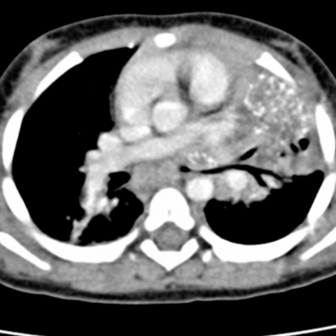
Chest computed tomography
Chest computed tomography