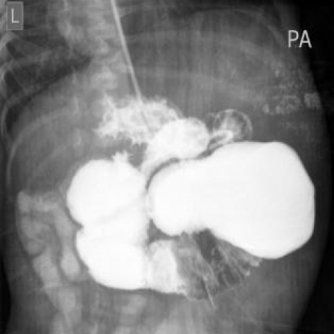
Figure 1
Abdominal radiograph-Erect postion

Dilated air filled stomach and proximal bowel loops with no evidence of air in the distal loops, nasogastric tube is seen inside the stomach
Paediatric radiology
Case TypeClinical Cases
AuthorsDr. Jamshid Sadiqi1, Dr. Najibullah Rasouly1, Dr. Homayoon Ghairatmal2, Dr. Hidayatullah Hamidi1, Dr. Shirazuddin Quraishi2, Dr. Reshad Faizi1, Dr. Nawaz Nasery1
6 days, female
[1] Berrocal, T., Lamas, M., Gutiérrez, J., Torres, I., Prieto, C., & del Hoyo, M. L. (1999) Congenital anomalies of the small intestine, colon, and rectum 1. Radiographics 19(5): 1219-1236.
[2] Louw, J. H. (1966) Jejunoileal atresia and stenosis. Journal of Pediatric Surgery 1(1): 8-23.
[3] Leonidas, J. C., Amoury, R. A., Ashcraft, K. W., & Fellows, R. A. (1976) Duodenojejunal Atresia with “Apple-Peel” Small Bowel: A Distinct Form of Intestinal Atresia 1. Radiology 118(3): 661-665.
[4] Manning, C., Strauss, A., & Gyepes, M. T. (1989) Jejunal atresia with'apple peel'deformity. A report of eight survivors. Journal of perinatology: official journal of the California Perinatal Association 9(3): 281-286.
[5] Waldhausen, J. H., & Sawin, R. S. (1997) Improved long-term outcome for patients with jejunoileal apple peel atresia. Journal of pediatric surgery 32(9): 1307-1309.
[6] Seashore, J. H., Collins, F. S., Markowitz, R. I., & Seashore, M. R. (1987) Familial apple peel jejunal atresia: surgical, genetic, and radiographic aspects. Pediatrics 80(4): 540-544.
[7] Peetsold, M. G., Ekkelkamp, S., & Heij, H. A. (2004) Late presentation of a duodenal web in a patient with situs inversus and apple peel jejunal atresia. Pediatric surgery international 20(4): 301-303.
| URL: | https://www.eurorad.org/case/13602 |
| DOI: | 10.1594/EURORAD/CASE.13602 |
| ISSN: | 1563-4086 |
This work is licensed under a Creative Commons Attribution-NonCommercial-ShareAlike 4.0 International License.

Abdominal radiograph-Erect postion
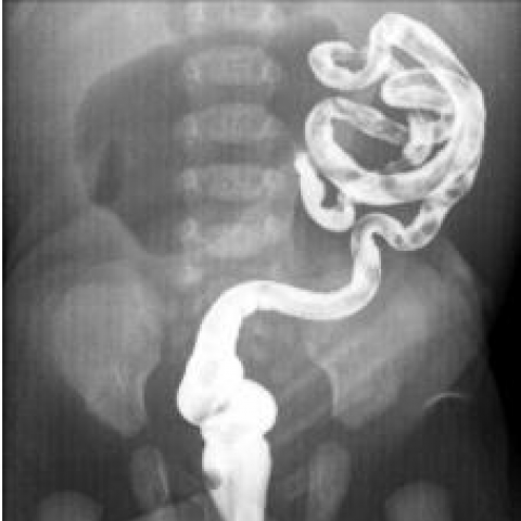
Upper GI study
Lower GI study
Lower GI study
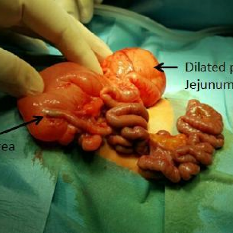
Surgery Image