Figure 1
Axial post-contrast CT

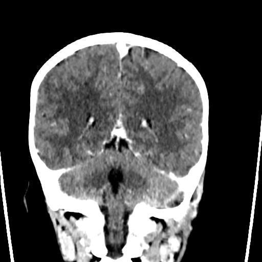
Axial contrast-enhanced CT of the head demonstrates filling defect with surrounding enhancement in the left sigmoid sinus and Bezold abscess.
Paediatric radiology
Case TypeClinical Cases
AuthorsVincent Leung, Vishal Bhalla, Nadir Khan
7 years, male
[1] Ross AK, Croft PR, Collins M (1988) Incident of acute otitis media in infants in a general practice. J R Coll Gen Pract 38(307):70-2 (PMID: 3204570)
[2] Kvaerner KJ, Bentdal Y, Karevold G (2007) Acute mastoiditis in Norway: no evidence for an increase. Int J Pediatr Otorhinolaryngol 71(10):1579-83 (PMID: 17707917)
[3] Vazquez E, Castellote A, Piqueras J, Mauleon S, Creixell S, Pumarola F, Figueras C, Carreño JC, Lucaya J (2003) Imaging of complications of acute mastoiditis in children. Radiographics 23(2):359-72 (PMID: 12640152)
[4] Castillo M, Albernaz VS, Mukherji SK, Smith MM, Weissman JL (1998) Imaging of Bezold's abscess. Am J Roentgenol 171(6):1491-5 (PMID: 9843276)
[5] Wang L, Leach J (2013) Intracranial complications of sinusitis and mastoiditis in children: imaging spectrum. RANZCR ASM 2013 Scientific Exhibit
[6] Go C, Bernstein JM, de Jong AL, Sulek M, Friedman EM (2000) Intracranial complications of acute mastoiditis. Int J Pediatr Otorhinolaryngol 52(2):143-8 (PMID: 10767461)
[7] Pang LH, Barakate MS, Havas TE (2009) Mastoiditis in a paediatric population: a review of 11 years experience in management. Int J Pediatr Otorhinolaryngol 73(11):1520-4 (PMID: 19758711)
[8] Minks DP, Porte M, Jenkins N (2013) Acute mastoiditis--the role of radiology. Clin Radiol 68(4):397-405 (PMID: 22980753)
[9] Wong AM, Zimmerman RA, Simon EM, Pollock AN, Bilaniuk LT (2004) Diffusion-weighted MR imaging of subdural empyemas in children. Am J Neuroradiol 25(6):1016-21 (PMID: 15205140)
| URL: | https://www.eurorad.org/case/13567 |
| DOI: | 10.1594/EURORAD/CASE.13567 |
| ISSN: | 1563-4086 |
This work is licensed under a Creative Commons Attribution-NonCommercial-ShareAlike 4.0 International License.

Axial post-contrast CT
Coronal post-contrast CT
Axial CT bone windows
Axial T2 weighted MRI
Diffusion-weighted MRI
Maximum intensity projection of MR venogram
Post-contrast T1 weighted MRI