Clinical History
A 25-year-old male patient with haematochezia was evacuated from the airport to our emergency room. He was haemodynamically unstable with haemoglobin of 3.8g/dL. Previous clinical data included digestive tract bleeding since two weeks followed by a colonoscopy with rectal polyp excision (pathology results unavailable) and a recent diagnosis of HIV infection.
Imaging Findings
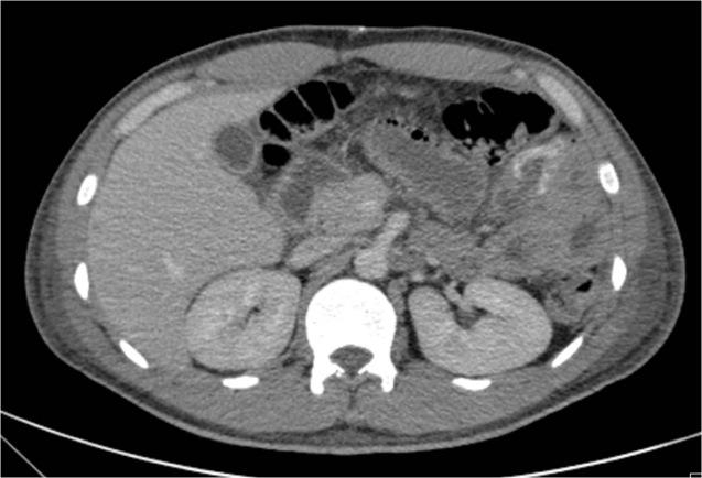
After stabilization and considering the history of previous rectal polyp excision (with the possibility of procedure complications taken into account), an angio-CT of the abdomen and pelvis was requested as primary evaluation, depicting a blush of contrast within the wall of a proximal small bowel loop, visible in the arterial phase and more conspicuous in the portal venous phase (Fig. 1 and Fig. 2). A vascular malformation was suspected as cause of the haemorrhage and the patient underwent immediate laparotomy. Intraoperatively, a small rigid nodule was discovered on the wall of a jejunal loop and a surgical incision was performed proximally to the lesion, allowing for an enteroscopy. In the middle jejunum, a 1 cm-pseudopolypoid lesion with surface hyperaemia and fresh adherent clot was found (Fig. 3). A segmental enterectomy was performed with uneventful postoperative recovery. The pathology analysis of the resected specimen revealed aspects consistent with Dieulafoy’s lesion (Fig. 4).
Discussion
Background
Although it had been first identified by Gallard, it was Paul Georges Dieulafoy who, in 1898, described the lesion in more detail, naming it “exulceratio simplex”. [1, 2] Also called “calibre persistent artery” [3], it corresponds to a histologically normal large and tortuous artery coursing in the submucosa with a maintained calibre of 1 to 3 mm [2, 4], which failed to taper distally. This artery usually protrudes through a small mucosal defect of a gastrointestinal tract organ [2, 4]. The most frequent location of Dieulafoy’s lesion is the stomach, but it can also occur more rarely in the oesophagus, duodenum, jejunum-ileum and colon. [2] The pathophysiologic mechanism of rupture and bleeding is still not completely clarified, although agreement seems to exist on the necessity of a mucosal insult (whether from erosion or ischaemia) that uncovers the silent vascular abnormality. [1]
Clinical Perspective
The usual clinical presentation of Dieulafoy’s lesion comprises intermittent and massive gastrointestinal bleeding, including melaena, haematemesis, haematochezia or a combination of more than one of these [5], depending on the location of the lesion. The diagnosis can be challenging as the lesion is inapparent in periods other than the ones with active haemorrhage. [2]
Imaging Perspective
Endoscopic studies (upper endoscopy, push enteroscopy, intraoperative enteroscopy and colonoscopy, depending on the location of the lesion) are useful for the diagnosis of the condition, but as previously said the discontinuous behaviour of the haemorrhage may pose diagnostic problems. Wireless capsule endoscopy has also been successfully applied. [1] The endoscopic criteria for the diagnosis of Dieulafoy’s lesion include: active haemorrhage from a less than 3 mm mucosal defect; visualization of a vessel protruding from a discrete defect or normal mucosa and/or fresh clot adherent to a defect of normal mucosa. [5]
The CT findings of Dieulafoy’s lesion comprise the detection of an enlarged submucosal vessel that can appear linear, tortuous or as a non-specific blush of contrast medium [2], as in our case.
Although there are no specific diagnostic findings at angiography, the diagnosis is suggested by detection of a tortuous and ectatic artery, otherwise normal appearing, associated with contrast extravasation. [1]
Outcome
In general terms, endoscopy has proven to be effective for the diagnosis and treatment of the majority of cases, with interventional angiography as a therapeutic alternative and surgical intervention reserved for the cases of failure with the previous techniques. Naturally, the treatment options will depend on the presentation of the disease, the location of the lesion and the available expertise. [1]
Differential Diagnosis List
Dieulafoy’s lesion of the jejunum
Arteriovenous malformations
Telangiectasias
Vascular tumours
Final Diagnosis
Dieulafoy’s lesion of the jejunum