[1]
Florin, Todd a Zaoutis, Theoklis E Zaoutis, Lisa B (2008) Beyond cat scratch disease: widening spectrum of Bartonella henselae infection. Pediatrics 121; e1413-e1425 (PMID: 18443019)
[2]
Rohr, Aaron Saettele, Megan R. Patel, Suchit A. Lawrence, Charles A. Lowe, Lisa H. (2012) Spectrum of radiological manifestations of paediatric cat-scratch disease. Pediatric Radiology 42; 1380-1384 (PMID: 22797536)
[3]
Danon, O. Duval-Arnould, M. Osman, Z. Boukobza, B. Kazerouni, F. Cadranel, J. F. (2000) Hepatic and splenic involvement in cat-scratch disease: Imaging features. Abdominal Imaging 25; 182-183 (PMID: 10675462)
[4]
García, Juan C. Núñez, Manuel J. Castro, Begoña Fernández, Jesús M. Portillo, Aránzazu Oteo, José a. (2014) Hepatosplenic Cat Scratch Disease in Immunocompetent Adults. Medicine 93; 267-279 (PMID: 26576306)
[5]
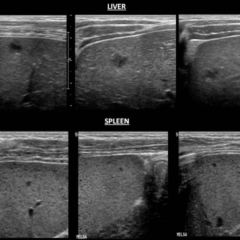
Rappaport, D. C. Cumming, W. A. Ros, P. R. (1991) Disseminated hepatic and splenic lesions in cat-scratch disease: Imaging features. American Journal of Roentgenology 156; 1227-1228 (PMID: 2028873)
[6]
Melville, D. M. Jacobson, J. a. Downie, B. Biermann, J. S. Kim, S. M. Yablon, C. M. (2015) Sonography of Cat Scratch Disease. Journal of Ultrasound in Medicine 34; 387-394 (PMID: 25715359)
[7]
Fouch, Brandy Coventry, Susan (2007) A case of fatal disseminated Bartonella henselae infection (cat-scratch disease) with encephalitis. Archives of Pathology and Laboratory Medicine 131; 1591-1594 (PMID: 17922599)
[8]
Marienfeld, Carla B. DiCapua, Daniel B. Sze, Gordon K. Goldstein, Jonathan M. (2010) Expressive aphasia as a presentation of encephalitis with Bartonella henselae infection in an immunocompetent adult. Yale Journal of Biology and Medicine 83; 67-71 (PMID: 20589186)
[9]
Rondet, B. Sarret, C. Lacombe, P. Rouveyrol, F. Chenel, C. Romaszko, J.-P. Labbé, A. (2012) Atteintes neurologiques à Bartonella henselae : à propos de 2 cas pédiatriques. Archives de Pédiatrie 19; 823-826 (PMID: 22749487)
[10]
Schmalfuss, Ilona M. Dean, Cooper W. Sistrom, Chris Bhatti, M. Tariq (2005) Optic neuropathy secondary to cat scratch disease: Distinguishing MR imaging features from other types of optic neuropathies. American Journal of Neuroradiology 26; 1310-1316 (PMID: 15956488)
[11]
HA, Carithers AM, Margileth (1991) Cat-scratch disease: Acute encephalopathy and other neurologic manifestations. American Journal of Diseases of Children 145; 98-101 http://dx.doi.org/10.1001/archpedi.1991.02160010104026 (PMID: 1845921)