Clinical History
A 42-year-old man presented 5 times to the emergency department with acute urinary retention. He had a previous history of renal colic.
Blood tests showed normal renal function and elevated WBC count. Urinalysis revealed microhaematuria.
He was discharged with a Foley catheter and medicated for prostatitis/urethritis and with α adrenergic receptor blocker.
Imaging Findings
Due to treatment failure, unenhanced CT of the abdomen and pelvis was performed, revealing two impacted urethral stones in the posterior urethra, with smooth contour and high density. The two stones combined measured at least 1.6 cm in the long axis.
The urinary bladder was weakly distended due to the presence of the Foley catheter, adequately positioned.
In the urinary bladder another smaller stone was noted, with smooth contours and high density.
There was mild homo-lateral ureterohydronephrosis associated.
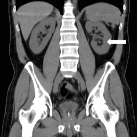
In the left kidney another two non-obstructive foci of nephrolithiasis were shown.
Discussion
A. Nephrolithiasis is a common disease. However urethral stones are rare and account for only 0.3-2% of all urinary tract stones. [1, 3, 4, 5]
They occur almost exclusively in men due to urethral anatomy, with a longer and more tortuous urethra in males. [1, 5] The calculus impacts more frequently in the posterior urethra, at prostatic or proximal membranous urethra, as this portion is non-distensible and the lumen is comparatively small. [1, 2]
More frequently, secondary or migratory stones form in the upper urinary tract and descend into the urethra. Less often, primary or native stones form in situ, secondary to urethral pathology such as strictures and diverticula. [1, 4, 5]
B. The most frequent presentation of an impacted urethral stone is acute urinary retention, but it can also be associated with irritative and/or obstructive symptoms and severe pain. Symptoms depend on the precise location of impaction and on the site of origin of the stone. [4, 5] Impacted stones in the posterior urethra produce perineal pain and in the anterior urethra penile pain. [1, 2] Secondary stones generally present with acute onset of symptoms and primary are usually associated with long history of lower tract symptoms.
Failure to diagnose and remove the impacted urethral stone can lead to post-obstructive renal failure, long-term urethral damage, urethrocutaneous fistulas, incontinence and impotence. [1]
Diagnosis of urethral stones can be challenging, as symptoms often are nonspecific and not every imaging modality for nephrolithiasis includes the lower urinary tract. [1]
C. Imaging plays a key role in diagnosis. The diagnosis of radiopaque calculi can be confirmed by plain radiography, as long as the urethra is also depicted. Ultrasonography can aid in the detection of urethral calculi, especially in radiolucent calculi. [1, 2] Unenhanced CT has become the gold standard for the evaluation of nephrolithiasis and has excellent sensitivity and specificity. As it usually includes the pelvis and therefore the lower urinary tract, all urethral stones should be identified. [1] Obstructive calculus generally have to be larger than 1 cm in diameter. [1, 5]
D. The treatment of choice is catheterization of the urethra or supra-pubic cystostomy followed by endoscopic management of such a stone.
E. In young and middle-aged males, especially those with history of nephrolithiasis, acute urinary retention is commonly caused by an urethral stone. [1, 2] CT urography generally includes the pelvis and when a urethral stone is present, it should be promptly identified. [1]
Differential Diagnosis List
Urethral calculi
Bone fragment
Strange body
Pelvic heterotopic ossification
Scrotal calculi