Figure 1
Initial contrast-enhanced multidetector CT at diagnosis

Axial (a,b in craniocaudal order) and coronal (c) postcontrast images showed homogeneously enhancing splenomegaly, absent splenic vein with enlarged tortuous perigastric venous collaterals (arrows) and gastric wall varices (thin arrows in b).

Axial (a,b in craniocaudal order) and coronal (c) postcontrast images showed homogeneously enhancing splenomegaly, absent splenic vein with enlarged tortuous perigastric venous collaterals (arrows) and gastric wall varices (thin arrows in b).

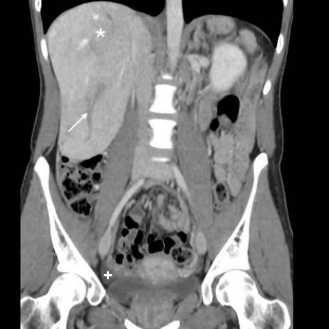
The enlarged spleen showed a focal hypovascular region (arrowhead) at the upper pole, consistent with limited infarct. Note enlarged tortuous perisplenic, perigastric and periduodenal venous collaterals (arrows).

Panoramic coronal maximum intensity projection (MIP) reconstruction showed patent portal and superior mesenteric veins, absent splenic vein, extensive venous collaterals (arrows) and gastric wall varices (thin arrows).

Axial thick-slab maximum intensity projection (MIP) reconstruction showed patent portal vein, absent splenic vein, venous collaterals (arrow) and gastric wall varices (thin arrows).