


Figure 1
Sagittal T2 Cervicothoracic spine
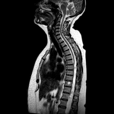
Sagittal T2 Cervicothoracic spine.
Neuroradiology
Case TypeClinical Cases
AuthorsW. Al-Obaydi, S. Patel.
56 years, female
[1] Lee H-J et al (2001) Symptomatic spinal intradural arachnoid cysts in the pediatric age group: description of three new cases and review of the literature. Pediat Neurosurg 35:181–7. (PMID: 11694795)
[2] Osborn AG (1994) Tumours, cysts, and tumour like lesions of the spine and spinal cord. diagnostic neuroradiology. St Louis, MO: Mosby Year Book, Inc., 876–918
[3] Kazan S, Ozdemir O, Akyuz M, Tuncer R (1999) Spinal intradural arachnoid cysts located anterior to cervical spinal cord: report of two cases and review of literature. J Neurosurg (Spine 2) 91:211–5 (PMID: 10505507)
[4] Teng P, Rudner N (1960) Multiple arachnoid diverticula. Arch Neurol 2: 348–356 (PMID: 13837415)
[5] Osenbach RK, Godersky JC, Traynelis VC et al (1992) Intradural extramedullary cysts of the spinal canal: clinical presentation, radiographic diagnosis, and surgical management. Neurosurgery 30:35–42 (PMID: 1738453)
[6] Masuzawa H, Nakayama H, Shitara N, Suzuki T (1981) Spinal cord herniation into a congenital arachnoid cyst causing Brown-Séquard syndrome: case report. J Neurosurg 55:983–986 (PMID: 7299475)
[7] McCrum C, Williams B (1982) Spinal extradural arachnoid pouches: report of two cases. J Neurosurg 57:849–852 (PMID: 7143073)
[8] Kjos BO, Brant-Zawadzki M, Kucharaczyk W, Kelly WM, Norman D, Newton TH. (1985) Cystic intracranial lesions: magnetic resonance imaging. Radiology 155:363–9. (PMID: 3983386)
[9] Lolge S, Chawla A, Shah J, Patkar D, Seth M. (2004) MRI of spinal intradural arachnoid cyst formation following tuberculous meningitis. The British Journal of Radiology 77 (2004), 681–684 (PMID: 15326049)
[10] Khosla A, Wippold FJ. (2002) CT myelography and MR imaging of extramedullary cysts of the spinal canal in adult and pediatric patients. AJR Am J Roentgenol 178 (1): 201-7. (PMID: 11756120)
[11] Brugières P, Malapert D, Adle-biassette H et-al. (1999) Idiopathic spinal cord herniation: value of MR phase-contrast imaging. AJNR Am J Neuroradiol 20 (5): 935-9 (PMID: 10369369)
| URL: | https://www.eurorad.org/case/12636 |
| DOI: | 10.1594/EURORAD/CASE.12636 |
| ISSN: | 1563-4086 |
Sagittal T2 Cervicothoracic spine
Sagittal T1 Cervicothoracic spine
Sagittal T2 Lumbosacral spine
Axial T2
Axial T2


