Figure 1
Teaching Case
SectionUroradiology & genital male imaging
Case TypeClinical Cases
AuthorsCatarina Costa, Maria Helena Valentim
Connected authors
81 years, female
81-year-old woman admitted at our emergency department after being found unconscious at home. Apyretic, slightly dehydrated. Laboratory tests showed slight anaemia (11.4 mg/dL) and aggravation of chronic renal disease. No leukocytosis. Recent hospitalization due to urinary tract infection by E. Coli, treated with Amoxicillin and Clavulanic acid.
Emphysematous cystitis
Emphysematous pyelonephritis
Vesicocolic or vesicovaginal fistulas
First abdominal plain film performed when the patient was admitted to the emergency department showed no pathological findings, but the pelvis was not included in the image.
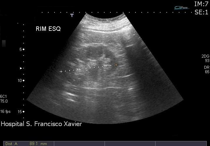
Due to laboratory aggravation of chronic renal function, renal ultrasound was performed, showing slight decrease in kidney length and parenchymal thickness, diffuse increase in cortical echogenicity and corticomedullary differentiation and slight hydronephrosis, confirming chronic renal disease. When bladder evaluation was performed, the posterior wall was ill-defined due to dirty shadowing artefact, and air inside the wall was immediately suspected. Second abdominal plain film, oriented to the pelvis, showed curvilinear areas of increased radiolucency delimitating inner bladder contour.
Pelvic CT was then performed, demonstrating diffuse wall thickness and corroborating the suspicion of air within the wall. Slight increase in mesenteric fat density was also found, probably reactive. Urethral catheter balloon was correctly positioned in the bladder lumen.
Emphysematous cystitis is an uncommon but severe disease, consisting of acute infection of the bladder mucosa and underlying musculature, produced by gas fermenting bacterial and fungal pathogens.
Patients may complain about dysuria, increased urinary frequency, haematuria and rarely pneumaturia. However, clinical presentation may be subtle and nonspecific, and may not represent the inflammatory nature of this disorder. This way, final diagnosis frequently relies on imaging findings [1].
The most frequently isolated pathogen is E. coli [2], but many other organisms have been reported such as Enterobacter aerogens, Klebsiella pneumonia, Proteus mirabilis, Staphylococcus aureus, Streptococci, Clostridium perfringens, and Candida albicans. Less commonly, it may have noninfectious causes such as bladder instrumentation, vesicocolic or vesicovaginal fistulas and trauma.
Once emphysematous cystitis is clinically suspected, imaging evaluation usually starts with conventional radiology. Plain films characteristically show curvilinear or mottled areas of increased radiolucency inside the urinary bladder, delineating its contour. Intraluminal gas can be detected as a non-static air-fluid level, and when adjacent to the non-dependent mucosal surface, it may have a characteristic cobblestone or “beaded necklace” appearance, which represents the irregular thickening produced by submucosal blebs [3].
US may also support the diagnosis, showing diffuse bladder wall thickening and increased echogenicity, and in some cases air may be seen in the form of hyperechoic echoes with posterior dirty acoustic shadowing.
CT allows early detection of intraluminal and intramural gas with high sensitivity, enabling also detection of some noninfectious causes of intraluminal gas, such as, for example, the presence of fistulas.
Our patient has some risk factors that increase susceptibility to emphysematous cystitis, namely being a female, poorly controlled diabetic, with a background of previous recurrent urinary tract infections. The fact that she had no fever or leukocytosis may be attributable to the antibiotic treatment she was receiving for a previous urinary infection.
Imagiologic evaluation showed all the typical findings of the disease, reported previously.
Emphysematous cystitis may be a potentially life-threatening condition, especially in older patients with comorbidities, therefore demanding for aggressive treatment with parenteral antibiotics and bladder drainage.
Surgical intervention is rarely needed, except when an anatomical abnormality is present (e.g. obstruction or a stone) [4].
References
[1] David E. Grayson, Robert M. Abbott, Angela D. Levy, Paul M. Sherman (2002) Emphysematous Infections of the Abdomen and Pelvis: A Pictorial Review. Radiographics 22: 543-561 (PMID: 12006686)
[2] Anil A. Thomas, Brian R. Lane, Arun Z. Thomas, Erick M. Remer, Steven C. Campbell, Daniel A. Shoskes (2007) Emphysematous cystitis: a review of 135 cases. BJU International 100(1):17-20 (PMID: 17506870)
[3] Alper Eken, MD, FEBU* and Ergun Alma, MD (2013) Emphysematous cystitis: The role of CT imaging and appropriate treatment. Can Urol Assoc J 7(11-12):E754-6 (PMID: 24282470)
[4] Masayuki Amano and Taro Shimizu (2014) Emphysematous Cystitis: A Review of the Literature. Intern Med 53: 79-82 (PMID: 24429444)
Case information
| URL: | https://www.eurorad.org/case/11515 |
| DOI: | 10.1594/EURORAD/CASE.11515 |
| ISSN: | 1563-4086 |