CASE 11322 Published on 04.11.2013

Long oesophageal leiomyoma

Section

Chest imaging

Case Type

Clinical Cases

Authors

Daniel Ramos Andrade, Célia Antunes, Luís Curvo Semedo, Filipe Caseiro Alves

Medical Imaging Department and Faculty of Medicine,
University Hospital of Coimbra, Portugal;
Email:daramosandrade@gmail.com
Patient

65 years, male

Categories
Area of Interest Gastrointestinal tract ; Imaging Technique MR, Fluoroscopy
Clinical History
A previously healthy 65-year-old male patient presented with a five-year history of mild dysphagia and occasional nausea and vomiting. Physical examination and blood work were unremarkable. A barium swallow and a thoracic MRI were performed.
Imaging Findings
Barium swallow oesophagogram
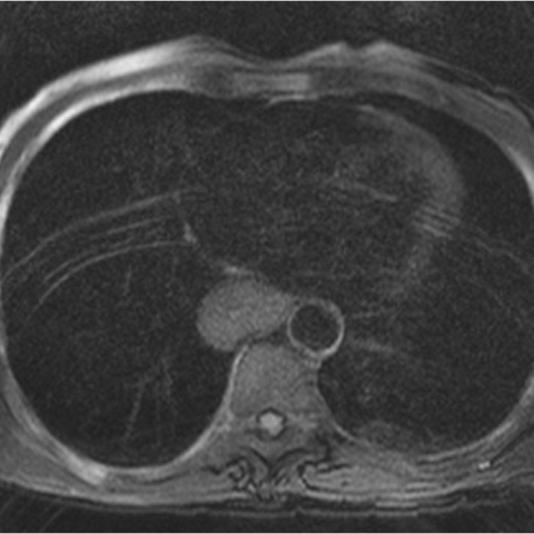
 showed a circumferential and symmetric filling defect, smoothly tapering, approximately 10 cm in length, localized at the mid and distal third of the oesophagus. The lesion had obtuse implantation angles and there was no irregularity of the overlying mucosa, which strongly favoured an extramucosal origin. There was weak distensibility throughout the lesion, which caused moderate obstruction to the flow of barium (Figure 1, 2).

Thoracic MRI was performed and demonstrated circumferential parietal thickening with soft tissue intensity, isointense relative to the skeletal muscle, in the mid third of the oesophagus, approximately 10 cm in length (Figure 4). There were no signs of invasion of the lung, other organs of the mediastinum or the thoracic spine (Figure 5). There was mild dilation of the proximal oesophagus (Figure 6, 7).
Discussion
Oesophageal leiomyoma is a rare benign tumour. It is the most common mesenchymal tumour of the oesophagus, unlike in the remainder of the gastrointestinal tract, where GISTs predominate. It is more common in men, between 20 and 50 years of age.
It is found more often in the lower two thirds of the oesophagus, which is consistent with the normal anatomical distribution of smooth muscle within the oesophageal wall.
It is usually an accidental finding but can be symptomatic (epigastric pain, dysphagia, pyrosis, oesophageal obstruction and reflux) when very large.
It consists of an encapsulated lesion of intersecting bands of muscle (spindle cells) and fibrous tissue. The cells have a bland appearance without nuclear pleomorphism, and there is little to no mitotic activity. Leiomyomas occasionally may contain dense areas of calcification, but cystic degeneration, necrosis, and ulceration almost never occur. The potential for malignant degeneration of leiomyomas is extremely small.
It habitually presents as a discrete, spherical or oval submucosal mass, ranging from 2 to 8 cm in size. Occasionally, it can encircle the oesophagus in a circumferential way and cause obstructive symptoms (as in our case). In about 5% of cases it can be multiple [1, 2, 3].

An oesophageal leiomyoma may appear as a posterior mediastinal mass on a chest radiography, with an abnormal azygoesophageal line.
On a barium meal, an oesophageal leiomyoma typically manifests as a smoothly marginated, round or lobulated intramural extramucosal mass, which typically forms right angles or slightly obtuse angles with the adjacent oesophageal wall.
Because oesophageal leiomyomas are submucosal lesions, conventional endoscopy is not a reliable diagnostic method.
Oesophageal ultrasound establishes either the muscularis mucosa or muscularis propria origin of the mass, without invasion of the mucosa or adventitia.
On CT examination, a homogeneous intramural mass or oesophageal wall thickening is found. Surrounding mediastinal fat and adjacent organs are not usually disrupted or invaded. It has a homogeneous iso or low attenuation before and after intravenous contrast administration.
MR examinations usually show isointensity or slight hyperintensity in T2-weighted images (in contrast with oesophageal carcinoma which is hyperintense) [4, 5].

Local enucleation is the recommended treatment choice for all symptomatic or larger than 5 cm oesophageal leiomyomas. When it involves the oesophagus in a diffuse fashion (like our case), a complete oesophagectomy is required. Resection is the only way to confirm that the tumour is not malignant. Barium meal follow-up is advised for smaller or asymptomatic lesions [5].
Differential Diagnosis List
Oesophageal leiomyoma
Oesophageal carcinoma
Oesophageal lymphoma
Oesophageal GIST
Oesophageal leiomyomatosis
Final Diagnosis
Oesophageal leiomyoma
Case information
URL: https://www.eurorad.org/case/11322
DOI: 10.1594/EURORAD/CASE.11322
ISSN: 1563-4086