Clinical History
A 72-year-old female patient presented with an incidentally found renal tumour of the left kidney. Histology proved a chromophobe oncocytoma and she underwent robotic assisted laparoscopic partial nephrectomy. The patient was now referred for worsening left flank pain over the past 12 months after surgery.
Imaging Findings
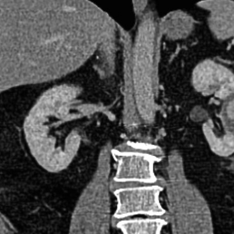
Contrast enhanced CT scan of the abdomen demonstrated a well circumscribed hypodense area of 7.5 x 5.5 cm containing an enhancing focus at the prior surgical site of the left kidney (Fig. 1). The central focus of 3 cm, showing vascular contrast dynamics in the arterial and delayed phase, allowed the diagnosis of a post-surgical renal artery pseudoaneursym (RAP) with surrounding haematoma. Angiography confirmed a 3.3 cm measuring pseudoaneurysm fed from a subbranch of a posterior segmental artery (Fig. 2). To minimise parenchymal loss, a very focal embolisation with 0.5 ml N-Butyl-2-Cyanoacrylate (NBCA) glue in a 1:2 dilution with iodised oil was performed close to the arterial lesion (Fig. 3). Subsequent contrast injection showed no further filling of the pseudoaneurysm. Follow-up CT at 45 days post-embolisation demonstrated the NBCA embolus in the posterior segmental artery with persistent lack of contrast filling and decreased size of the pseudoaneurysm (Fig. 4).
Discussion
A. BACKGROUND
Most RAP are of iatrogenic origin and related to minimal invasive procedures like biopsies, percutaneous nephrostoma or minimal invasive surgery. A pseudoaneurysm is caused by injury to a layer of the arterial wall, so that the blood flow is only contained by the remaining layers of the arterial wall or solely by surrounding tissue. Recent studies suggest a higher incidence of RAP with the advent of minimal invasive partial nephrectomies compared to open partial nephrectomy [1]. Angio-embolisation is the treatment of choice, since it combines reliable occlusion of the injured artery with the least amount of collateral nephron loss. Embolic agents used are coils, gelfoam or PVA particles. NBCA glue can become a preferred alternative agent in scenarios, where a very focal embolisation is desirable. Also, cost-efficiency may play a role, if deployment of multiple coils can be avoided.
B. CLINICAL PERSPECTIVE
Clinical presentations of RAP comprise one or several of three symptoms: Gross haematuria, flank pain and anaemia [1]. Any one of these warrants a thorough clinical assessment and work-up, since rupture of a pseudoaneurysm is a life-threatening event.
C. IMAGING PERSPECTIVE
ANGIOGRAPHY: Gold standard for diagnosis and therapy, direct contrast filling of the aneurysmatic sac after selective contrast injection can be seen. However, in the haemodynamically stable patient non-invasive modalities may be preferred.
CT ANGIOGRAPHY: Contrast filling of the outpouching sac adjacent to the renal artery with demonstration of wash-out on delayed phases. CE-CT is likely the non-invasive modality of choice, since it not only allows a positive diagnosis of a RAP, but also depicts possible differential diagnoses.
ULTRASOUND: The hallmark is a cystic structure communicating with the renal artery, and showing a “to and fro” waveform on Doppler termed “yin-yang sign”. Ultrasound can, however, be limited by body habitus and depend on examiner experience.
MR ANGIOGRAPHY: Flow void from the blood flow jet in the pseudoaneursym adjacent to the feeding artery can be visualised. [2]
D. OUTCOME:
Selective angio-embolisation is the established treatment of choice in the management of a RAP with coils being most frequently used. In select cases NBCA glue can, however, complement the armamentarium of peripheral embolisations. Use of NBCA requires a meticulous technique with thorough flushing of the delivery microcatheter with 5% dextrose to avoid preemptive polymerization of the glue [3].
E. TEACHING POINT: NBCA glue is a safe alternative embolisation in the treatment of RAP.
Differential Diagnosis List
Successful embolisation of a post-surgical renal artery pseudoaneursym using NBCA glue.
Arteriovenous malfomation
True aneurysm
Final Diagnosis
Successful embolisation of a post-surgical renal artery pseudoaneursym using NBCA glue.