Figure 1
First CT examination - Baseline and venous-phase acquisition

Baseline abdominal CT showing marked subcapsular renal fluid collection in the left perirenal space, displacing forwards the omolateral renal parenchyma; on its medial side in contact with omolateral psoas muscle and left wall of aorta.

Baseline abdominal CT showing marked subcapsular renal fluid collection in the left perirenal space, displacing forwards the omolateral renal parenchyma; on its medial side in contact with omolateral psoas muscle and left wall of aorta.

Baseline abdominal CT showing marked subcapsular renal fluid collection in the left perirenal space, displacing forwards the omolateral renal parenchyma; on its medial side in contact with omolateral psoas muscle and left wall of aorta.

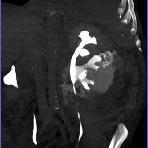
Venous-phase abdominal CT evaluation showing marked subcapsular renal fluid collection in the left perirenal space, within septations and with peripherical wall-enhancement.

Venous-phase abdominal CT evaluation showing marked subcapsular renal fluid collection in the left perirenal space, within septations and with peripherical wall-enhancement.

Venous-phase abdominal CT evaluation showing marked subcapsular renal fluid collection in the left perirenal space, within septations and with peripherical wall-enhancement.